Research by Amy Collins, PHD student funded by the William E Harker Foundation
A new human liver cancer culture model: precision cut liver slices (PCLS)
There is an urgent need to further understand the biology of liver cancer and develop new drugs for the treatment of this disease. Animal models and cell cultures do not mimic the human disease, because mice are not humans and human cells grown in 2D on plastic dishes do not recreate the complex 3D interactions between the cancer cells and the different types of liver cells found within the tumour-liver environment. Therefore, understanding the disease biology and developing effective drugs in these models can be challenging.
As part of our research, we are creating a new human liver cancer culture model, which does recreate the interactions between cancer cells, liver tissue and immune cells. This is achieved by adding small balls of liver cancer cells to very thin slices of human liver and then growing them in a special bioreactor. The cancer cells invade and grow within the liver slice and these slices can then be bathed in different drugs to ask if the drug can slow the growth of the cancer cells. We can also add immune cells to our liver cancer slice model to study the biology of these cells and ask if they affect cancer cell growth

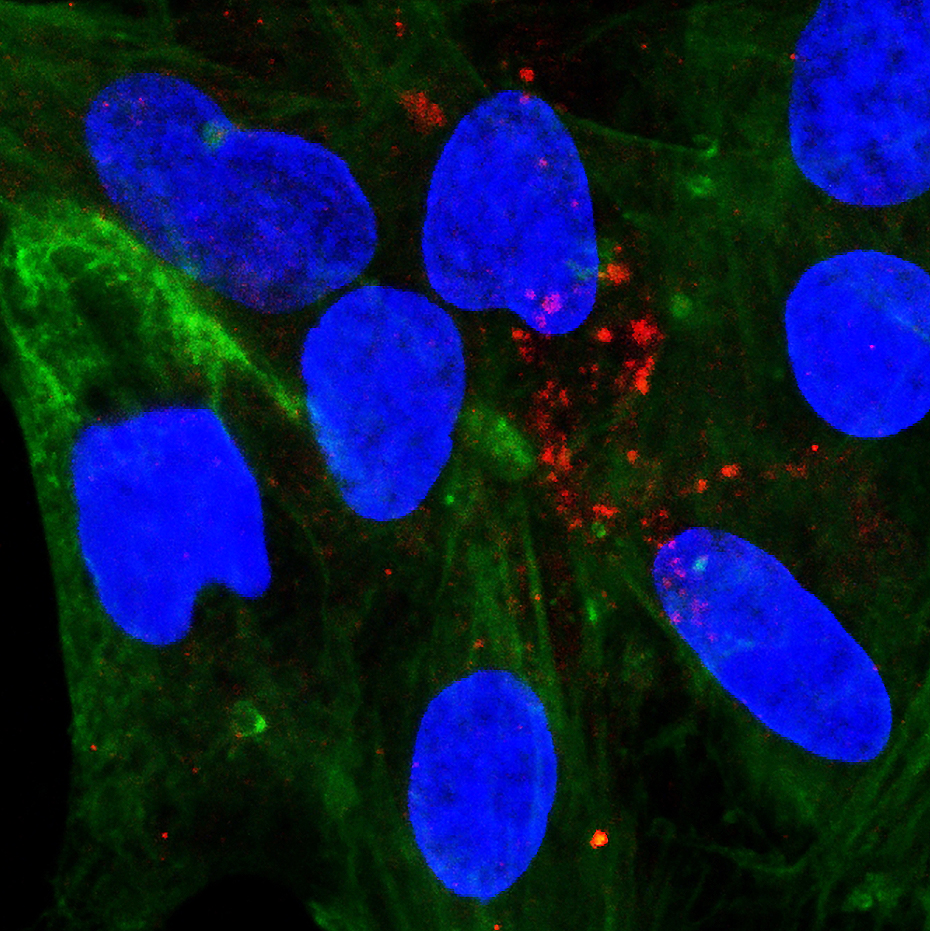
The image above shows human HCC cells (red) engrafted “entering” a human PCLS, nuclei (blue), with added immune cells (green). The orthogonal projection of sections from the top to the bottom of the slice (250 microns total) shows that the cancer cells penetrate throughout the slice, the immune cells labelled green, surround the tumour.
The image below shows Human HCC and IC – this is a top down view of the image above, and shows HCC cells (red) engrafted “entering” a human PCLS, nuclei (blue), with added immune cells (green).

Research by Maja Laszczewska, HUNTER Research Technician
Generating primary liver cancer cell lines
Maja Laszczewska has been successfully generating cell lines from surplus human primary liver cancer biopsy and resection tissues. She has been able to characterise the cells she has been growing using a variety of different cell-surface markers.
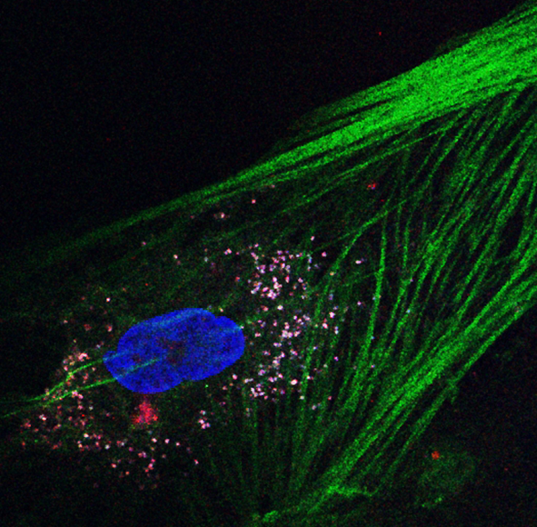
The image on the bottom left shows immunofluorescence staining in patient derived cancer associated fibroblasts (CAFs) for Sulfatase 2 (red) and alpha smooth muscle actin (green), and nuclei (blue).
The image on the bottom right shows immunofluorescence staining in patient derived cancer associated fibroblasts for Sulfatase 2 (red) and alpha smooth muscle actin (green), and nuclei (blue).
Sulfatase 2 (SULF-2) is an enzyme that is upregulated in hepatocellular carcinoma (HCC) and alpha smooth muscle actin (aAMA) is used to identify fibroblasts.
Exploration of ‘liquid biopsy’ biomarkers for HCC
Hepatocellular Carcinoma (HCC) is the fourth commonest cause of cancer death globally (attributed to viral hepatitis) and rising dramatically consequent to an obesity epidemic causing fatty liver and cancer. Screening tests for early detection in the at risk population are poor, with HCC typically detected at advanced stages. Traditional cytotoxic therapies are poorly tolerated (diseased/cirrhotic liver) and while medical therapy options are growing, their use is hampered by the lack of clinically useful biomarkers. Liver biopsy carries significant risk and even for diagnostic purposes is avoided if possible, limiting novel biomarker discovery/application.
Thus our team has a keen interest in developing liquid biopsy tools – focused on the tumours products (cells and nucleic acids – DNA, RNA) that are released into the circulation. Circulating tumour cells are quite rare in HCC patients and DNA analysis is promising mostly in terms of mutation analysis. Analysis of extracellular RNA (exRNA) is a highly attractive alternative to characterise the tumour, although challenging due to degradation of RNA in the circulation. We have optimised methods taking advantage of recent technology advances for analysing extracellular vesicles (EV) protected RNA. EVs play a crucial role in regulating the proliferation, immune escape, and metastasis of HCC. Thus we hypothesis that characterising the EV RNA ‘cargoes’ from HCC patients serum can provide useful biomarkers for HCC diagnosis and prognosis. Towards this we have secured 2 PhD studentships.
Ranjeet Singh: Alice Stephenson Bequest Fund PhD studentship. ‘Identification of ‘liquid biopsy’ predictive biomarkers in patients with advanced hepatocellular carcinoma’ (Sep 2022-Aug 2025)
Dame Margaret Barbour Foundation PhD studentship. Title: ‘Exploration of extracellular RNA (exRNA) from hepatocellular carcinoma patients serum for biomarker discovery’ (Sep 2022-Aug 2025)

Research by Dr Daniel Geh, clinical PhD student funded by the Newcastle Cancer Research UK (CRUK) Clinical Academic Training Programme
Neutrophils as potential therapeutic targets in hepatocellular carcinoma
Neutrophils are the most common white blood cell and are the immune systems first responders to infection and injury.
A growing body of evidence points to neutrophils as playing an important role in the development, progression and potential treatment of liver cancer.
My aim is to characterise in detail how neutrophils change in response to liver cancer and the effect this has on cancer progression. Using this information, I aim to then manipulate these changes as a potential therapeutic treatment. To achieve this, I am isolating and studying neutrophil populations from different organs in both patients and mouse models of liver cancer.